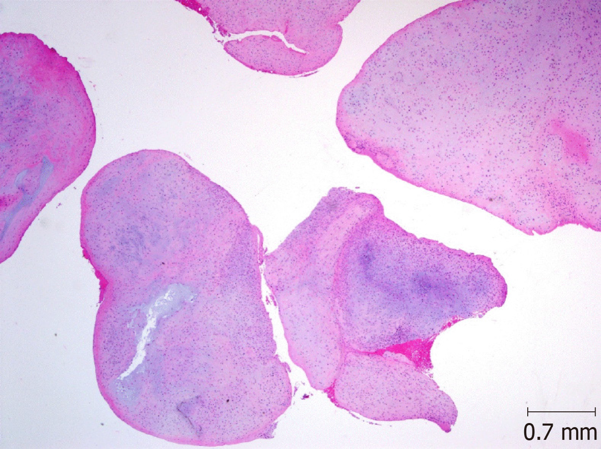
loose body histology
Third there is a zonal pattern of giant cells in the center which are. Loose or areolar connective tissue is found throughout the body wherever biologic packing material is needed.

Macro And Microscopic Findings Of The Loose Body A Extracted Loose Download Scientific Diagram
Dense regular connective tissue.

. Lets identify the thick and thin skin histology slides under a light microscope. You need to choose an epithelium optimized for an animal that lives in a sterile aquarium. This loose fibrous tissue is widely distributed in the body where it provides strength elasticity and support to neighboring tissues.
This is a section of areolar loose connective tissue from a non-adipose region of the subcutaneous layer of the integument. Lung histology classification the tissues of the lung derive from endoderm they are grouped together with other. Respiratory portion progresses from ciliated cuboidal epithelium to.
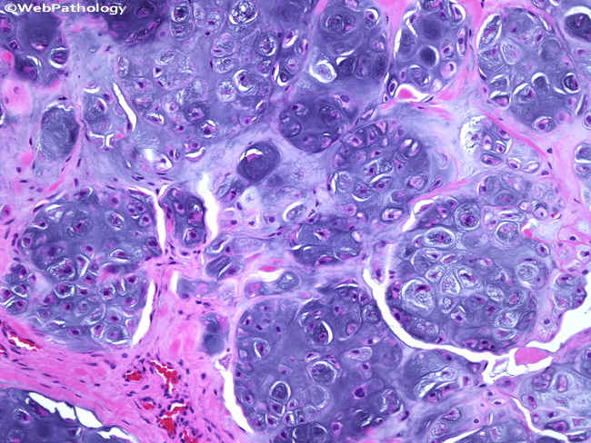
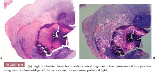
Three different types of cartilage were identified in the nidus of a loose body--articular. 3 loose bodies due to joint surface disintegration. 2 loose bodies due to osteochondral fracture.
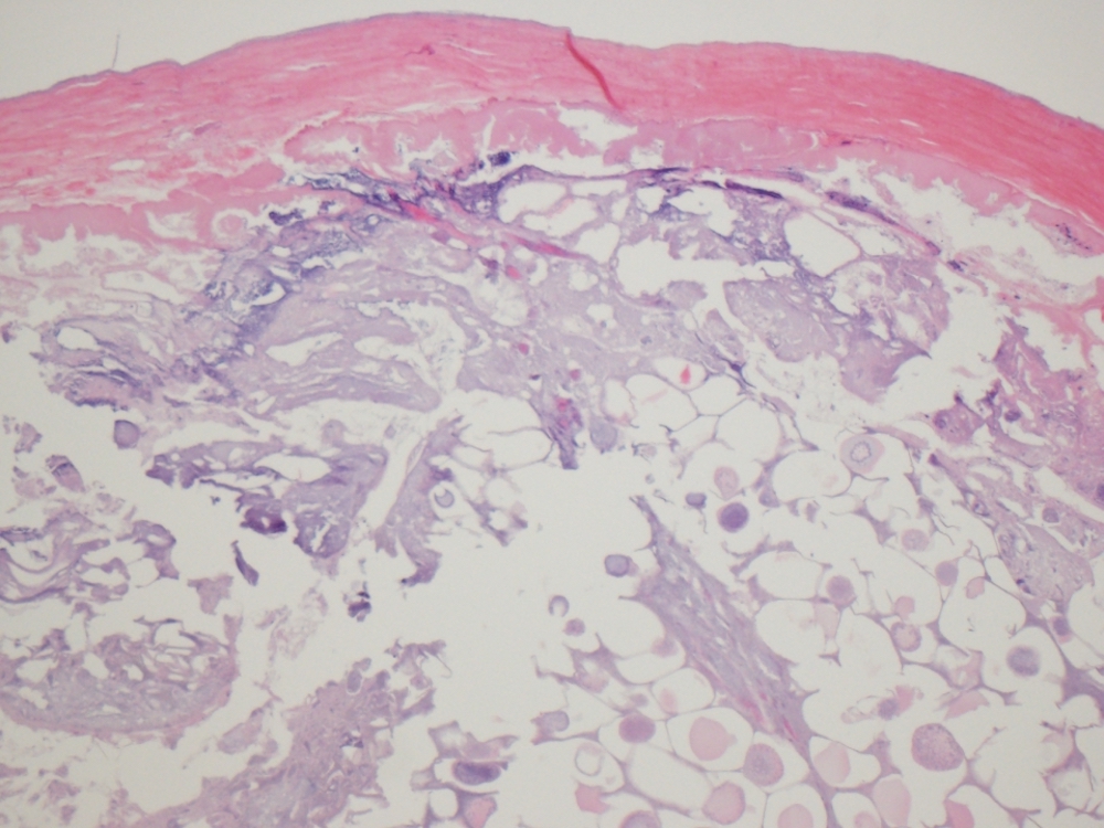
Dense irregular connective tissue. It is a. The provided tissue section shows two distinct layers the epidermis and dermis.
First talk about the thin skin microscope slide identification. At a histological level both the heart and blood vessels consist of three layers. 1 loose bodies due to synovial osteochondromatosis.
Neutrophils and macrophages are also present and both are discussed below. We histologically examined 84 loose bodies and 9 related lesions synovial membrane nodules surgically removed from 24 joints of 24 patients with osteoarthrosis. Loose areolar connective tissue Loose areolar CT is characterized by relatively loosely arranged collagen and elastic fibers.
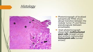
This region is also a loose irregular connective tissue but can be so extensively infiltrated by white blood cells and plasma cells that the supporting fibers and ground substance are obscured. Learn vocabulary terms and more with flashcards games and other study tools. Areas of multinucleated giant cells are admixed with fibrous tissue and reactive bone.
However few studies have focused on the functional histology of these layers and their relations with infertility in humans. Second the giant cells are admixed with extravasated RBCs. Loose CT or areolar tissue is the most widespread CT of the body.
First the giant cells are smaller than those of a typical giant cell tumor. The composition and organization of a particular connective tissue largely depends on its function. Presence of thin epidermis that lines with keratinized stratified squamous epithelium.
The histological structure is characterised by the functional nature of the structures. Histology - Areolar connective tissue View Related Images. It is characterized by an abundance of ground substance plus thin and relatively few fibres and cells Fig.
The main cellular elements are fibroblasts and a smaller amount of adipocytes. Muscular layer - smooth muscle in the blood vessels cardiac muscle myocardium in the heart. The 26 osteochondral loose bodies type II.
Histology of the lung is the study of the microscopic structure of the lung. It is well vascularized and highly cellular with a large proportion of matrix. It is highly cellular containing fibroblasts immune cells eg mast cells macrophages T cells and endothelial cells capillaries.
Endothelial layer - epithelial tissue formed by simple squamous endothelial cells. The 84 loose bodies included 48 chondral loose bodies type I 26 osteochondral loose bodies type II and 10 osseous loose bodies type III. The fibrous component varies in amount and orientation.
In the heart this layer is referred to as endocardium. There are several distinctive features of giant cell reparative granuloma. Loose CT can further be described as areolar or reticular.
A histopathological analysis of 119 surgically excised loose bodies revealed that the cases could be separated into three categories. Imagine you were hired by a pharmaceutical company to create membranes for genetically engineered animals that are different from all previous life forms found on earth. Plasma Cells Slide 29 small intestine HE WebScope.
Definition general Defined as infestation caused by body louse Pediculus humanus corporis Skin lesions due to direct bite hypersensitivity reaction and itching related excoriation Louse is also vector for other diseases such as.
Webpathology Com A Collection Of Surgical Pathology Images

A Rice Bodies Gross Specimen B Rice Bodies Microscopic Histology Download Scientific Diagram
Pvns Synovial Chondromatosis Loose Bodies
Histological Examination Of The Loose Bodies Fibrous Connective Tissue Download Scientific Diagram
Pathology Outlines Synovial Tenosynovial Chondromatosis

Histopathology Slide Of The Loose Body Showing Lobules Of Cartilage Download Scientific Diagram

Autoamputated Adnexa Presents As A Peritoneal Loose Body Fertility And Sterility

Cartilage Proliferation Is Diagnosed During Histopathologic Evaluation Download Scientific Diagram

Histologic Evaluation Of Osteochondral Loose Bodies And Repaired Tissues After Fixation Arthroscopy

Xmlinkhub

Figure 3 Arthroscopic Treatment Of A Case With Concomitant Subacromial And Subdeltoid Synovial Chondromatosis And Labrum Tear

Histological Slides Of Excised Synovial Chondromatosis Tissue At Power Download Scientific Diagram

A Loose Bodies Showing Endochondral Ossification In The Hyaline Download Scientific Diagram
Synovial Osteochondromatosis Of The Temporomandibular Joint A Case Report
Joints Basicmedical Key

Extracted Specimen A The Peritoneal Loose Body Measured 65 Mm Was Download Scientific Diagram

Pdf Uncalcified Synovial Chondromatosis In The Pisotriquetral Joint Semantic Scholar

9ws2smii C1zum
Pathology Outlines Infarcted Epiploic Appendages


Comments
Post a Comment